Human Anatomy and Physiology
The basic needs of the human race are food, water and oxygen. We take in food and water through the mouth and breathe in oxygen through the nose. The food is digested in the digestive system and is made absorbable by the secretions of pancreas, liver and intestinal glands. Oxygen taken into the respiratory passage reaches the lungs and enters the blood. For the proper functioning of the vital organs like heart, brain and other parts of the body, the absorbed food and oxygen must reach them. This is done by the circulatory system. The nerves stimulate the organs to function by carrying the commands given by the brain as electrical impulses. The excretory products formed in the various organs and the carbon dioxide released after the utilization of oxygen must be carefully separated from the tissues and sent out of the body. This is done by the excretory system. The body has five special sense organs - eye, ear, nose, tongue and skin to appreciate the external environment. All these systems cooperate with one another and function in coordination.
The eye is a vital sense organ of the human body. It is related to other organ systems in many ways. Hence in addition to knowing the structure and function of the eye, the anatomy (structure) and physiology (function) of other organ systems assumes paramount importance. This helps the OA to appreciate the importance of viewing the patient as a whole and not the eye alone.
The structure and function of the body systems are discussed in detail and they are:
- Cell
- Skeletal system
- Muscular system
- Cardio vascular system
- Respiratory system
- Nervous system and sense organs
- Excretory system
- Digestive system
- Endocrine system
- Reproductive system
1. Cellular System
Cells are the smallest units of our body. They are the basic units of our body.
The structure of a cell
The human body develops from a single cell. The single cells are derived from zygote. Zygote is formed by fusion of sperm and ovum. Zygote multiplies and develops into various structural and functional types of cells. The cell consists of a plasma membrane and cytoplasm.
Inside the plasma membrane, there are number of organelles floating in a watery fluid called cytoplasm. Organelles are small structures with highly specialized functions. Organelles include
- Nucleus
- Golgi bodies
- Mitochondria
- Ribosomes
- Endoplasmic reticulum
- Lysosomes
Plasma membrane
The thin layer of protein and fat that surrounds the cell is the cell membrane or the plasma membrane. The cell membrane is semi permeable, allowing some substances to pass into the cell and blocking others.
It consists of two layers with specialised functions.
- They give the cell its immunological identity.
- They can act as specific receptors for hormones and other chemical messengers.
- The plasma membrane has several enzymes embedded on its layers.
Organelles
Nucleus
Every cell (Fig. 3.1) in the body has a nucleus with the exception of mature erythrocytes (RBCs). The nucleus has a membrane similar to plasma membrane, called nuclear membrane. It has tiny pores through which certain substances pass between nucleus and cytoplasm. The nucleus contains chromosomes. The chromosomes have the body’s genetic material in the form of a large double chains of molecules of DNA (Deoxyribo Nucleic Acid). The nucleus controls the activities of a cell.
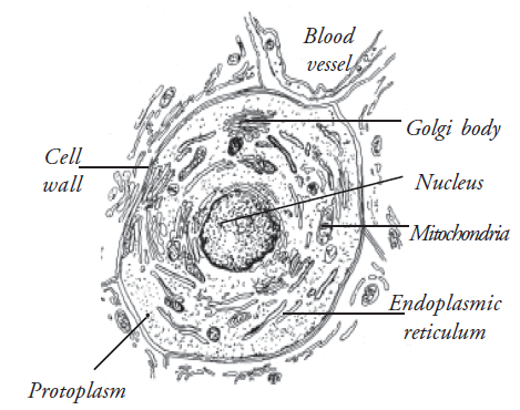
Mitochondria
These are sausage shaped structures in the cytoplasm. They are described as the “power house” of the cell. They are involved in cellular respiration. In cellular respiration energy is generated.
Ribosomes
These are tiny granules composed of RNA (Ribo Nucleic Acid) and protein. They synthesise proteins from amino acids.
Endoplasmic reticulum
They are a series of membranous canals in the cytoplasm. These are of two types, smooth and rough.
Golgi apparatus
This consists of stacks of closely folded flattened membranous sacs. They synthesise and export proteins.
Lysosomes
These are oval or spherical membrane bound bodies produced by the golgi bodies.
Cell division
There are two types of cell division: mitosis, meiosis.
Transfer of substances across cell membranes
Some substances cross plasma membranes by passing from a higher concentration on one side to a lower concentration on the other, without the use of energy. This is called diffusion (passive transport). Other types of transport are:
- Active transport
- Bulk transport
Tissues
The tissues of the body consist of large numbers of cells. They are classified according to the size, shape and functions of these cells. There are four main types of tissues, each of which has subdivisions. They are
- Epithelial tissue
- Connective tissue
- Muscle tissue
- Nervous tissue
Epithelial tissue
This group of tissues is found covering the body and lining cavities and tubes. It is also found in glands. The structure of epithelium is closely related to its functions which include:
- Protection of underlying structures
- Secretion
- Absorption
- Squamous
- Cuboidal
- Columnar
- Ciliated
- Stratified
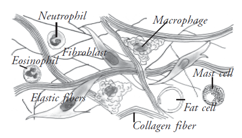
Connective tissue
It is found in all organs supporting the specialised tissue (Fig. 3.2). The different types of cells involved include:
- Fibroblasts
- Macrophages
- Plasma cells
- Mast cells
- Fat cells
Major functions of connective tissue are
- Binding and support
- Protection
- Transport
- Insulation
Types of connective tissue
- Areolar tissue
- Adipose tissue
- White fibrous tissue (collagen)
- Elastic tissue
- Reticular tissue
- Blood
- Lymphoid tissue
- Cartilage
- Bone
Muscle tissue
There are three types of muscle tissue:
- Striated, skeletal or voluntary muscle
- Non-striated, involuntary, visceral or smooth muscle
- Cardiac muscle
Nervous tissue
Two types of cells are found in the nervous system:
- Excitable cells
- Non-excitable cells
Organs
Organs are formed by a group of tissues. Each organ has its specific functions. e.g. Heart is made up of cardiac muscle which is a muscular tissue. It is the prime organ of systemic circulation.
Systems
A group of organs joins together and forms a system. We have the following systems in our body.
- Musculo-skeletal
- Circulatory
- Respiratory
- Nervous
- Endocrine
- Sense organs
- Excretory
- Reproductive
Anatomical terms
The anatomical position
This is the position assumed in all anatomical descriptions to ensure accuracy and consistency. The body is in the upright position with the head facing forward,
- The arms at the sides
- Palms of the hands facing forwards
- The feet together
- Toes directed forwards
Median plane
At anatomical position, the body is divided longitudinally into right and left halves, and this is median plane.
Medial and lateral
Any structure which is nearer to the midline is called medial. Any structure farther from the midline or at the side of the body is known as lateral.
Proximal and distal
These terms are used describing any structure that is long eg. the bones of the limbs. The proximal end of a bone is the one nearest to the point of attachment of the limb, and the distal end is farthest away.
Anterior
This indicates that the part being described is nearer the front of the body.
Posterior
This means that the part being described is nearer the back of the body.
Superior
This indicates a structure nearer the head.
Inferior
This indicates a structure farther away from the head.
Border
This is a ridge of bone which separates two surfaces.
Spine, spinous process or crest spine
This is a sharp elevation ridge or bone.
Trochanter, tuberosity and tubercle
These are roughened bony projections, usually for the attachment of muscles or ligaments. The different names are used according to the size of the projection. Trochanters are the largest and tubercles the smallest.
Fossa
This is a hollow or depression in a structure.
Foramen
This is a hole in a structure.
Bony sinus
This is a hollow cavity within a bone.
Meatus
This indicates a tube-shaped cavity within a bone.
Articulation
This is a joint between two or more bones.
Suture
This is the name given to an immovable joint, e.g. between the bones of the skull.
Articulating surface
This is the part of the bone which enters into the formation of a joint.
Septum
This is a partition separating two cavities.
Fissure or cleft
This indicates a narrow slit.
2. The Skeletal system
Skeletal system deals with bones and surrounding tissue. It forms the supporting framework of human body. This section includes
- Basic structure of a bone
- Classification of the bones
- Functions of the bones
- The different bones of the body
- Skull
- Vertebral column
- Thorax
- The appendicular skeleton
- Joints and movements
Basic structure of a bone
Bones are the hardest tissue in the body and when fully developed are composed of
Water - 20%
Organic material - 30- 40%
Inorganic material - 40- 50% (contains noncarbon)
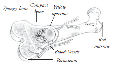
There are two types of bone tissue, compact and cancellous (Fig. 3.3).
A. Compact bone
- To the naked eye, compact bone appears to be solid
- These consist of a central canal
- This central canal is surrounded by concentric plates of bone cells, called the lamellae
- In these lamellae, there are lacunae or spaces, containing lymph and osteocytes
- These lamellae gives greater strength to the bone than a solid structure of the same size would
B. Cancellous bone
- To the naked eye, cancellous bone looks spongy
- The central canals are much larger than in compact bone
- There are fewer lamellae which gives a honey comb appearance
- Red bone marrow is always present in cancellous bone
Periosteum
- All the bones are almost completely covered by vascular fibrous membrane called " periosteum "
- This maintains the shape of the bone
- This gives attachment to muscles and tendons and protects bones from injury
Classification of bones
Bones are classified as
- Long
- Short
- Irregular
- Flat
- Sesamoid
Long bones
- They have a shaft (diaphysis) and two ends / epiphyses
- The shaft is composed of compact bone. It has a central cavity, called the medullary cavity. The medullary cavity contains " yellow bone marrow "
- The epiphyses consist of an outer covering of compact bone with cancellous bone inside
Short bones
- They are cuboidal in shape
- They have a peripheral compact bone and inner cancellous bone
- The carpal bones and tarsal bones are examples of short bones
Irregular, flat and sesamoid bones
- Irregular, flat and sesamoid bones are composed of a thin outer layer of compact bone with cancellous bone inside.
- These contains red bone marrow for example
Irregular bones form the - Vertebrae
Flat bones form the - Sternum, ribs
Sesamoid bones form the - Patella
Bone cells
There are two types of bone cells which are responsible for bone formation (Fig. 3.4).
- Osteoblasts
- Osteoclasts
Osteoblasts
These cells are present
- In deeper layers of periosteum
- In the immature bones
- At the ends of diaphysis
- At the site of fracture
As the bone develops, osteoblasts become trapped in the lacunae. They stop forming new bone at this stage and are called osteocytes. Nutrient artery of the bone arises from adjacent blood vessels. They supply the bone tissues.
Osteoclasts
Their function is resorption of bone to maintain optimum shape. This takes place at bone surfaces. A fine balance of osteoblasts and osteoclasts maintains a normal bone.
Functions of bones
- They provide frame work to the body
- Give attachment to muscles and tendons
- Helps in movements of the body
- Protects the internal organs
- Contains red bone marrow from which RBCs develop
- Provide a reservoir for calcium, phosphorous and fat
Bones of the skeleton
Bones of the skeleton are divided into two groups.
- Axial skeleton
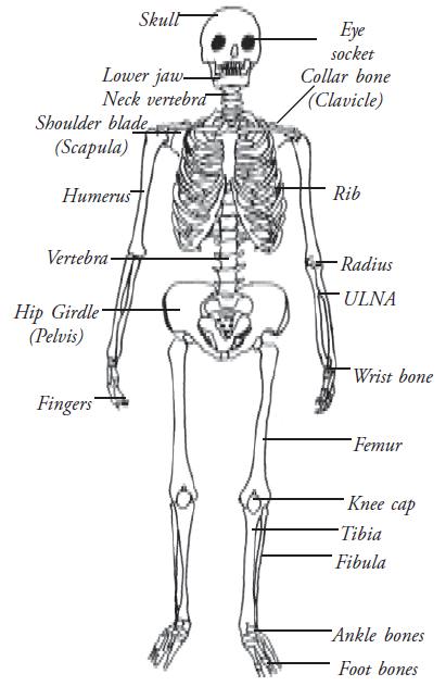
- Appendicular skeleton (Fig. 3.5)
Axial skeleton
This has the following parts
- Skull
- Vertebral column
- Thoracic cage
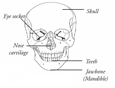
Skull
This rests on the upper end of vertebral column and has two parts 1. Cranium and 2. Face
Cranium
- 1 frontal bone
- 2 parietal bones
- 2 temporal bones
- 1 occipital bone
- 1 sphenoid bone
- 1 ethmoid bone
Face
- 2 zygomatic / cheek bones (Fig. 3.6)
- 2 maxilla
- 2 nasal bones
- 2 lacrimal bones
- 1 vomer
- 2 palatine bones
- 2 inferior conchae bones
- 1 mandible - (the only movable bone of the skull)
Hyoid bone
This is an horse shoe shaped bone lying above the larynx and below the mandible
Sinuses
Sinuses are cavities present in the bone and contains air. All sinuses communicate with the nose.
Functions of sinuses
- Gives resonance to the voice
- Helps in balancing the head by lightening the skull bones
The Vertebral column
The vertebral column consists of
- 7 Cervical vertebrae
- 12 Thoracic vertebrae
- 5 Lumbar vertebrae
- Sacrum (5 fused vertebrae)
- Coccyx (4 fused vertebrae)
The first cervical vertebra is called Atlas. The second cervical vertebra is called axis. The thoracic vertebrae are attached with the ribs. All the vertebrae are separated by intervertebral discs. When two adjacent vertebrae are viewed from the side, a foramen can be seen. Half of the wall is formed by the upper vertebra. The other half is formed by the lower vertebra.
There are inter vertebral foramen in each side, between adjacent vertebrae through which the spinal nerves emerge. The ligaments present in the vertebrae hold the vertebra together. It help to maintain the inter vertebral discs in position.
Functions of the vertebral column
- This gives a strong bony protection for the delicate spinal cord lying within it
- The intervertebral foramina act as passage for the spinal nerves, blood vessels, and the lymph vessels
- It supports the skull
- It allows a certain amount of movement
- The intervertebral discs act as a shock absorber protecting the brain
Thoracic cage
The bones of the thoracic cage are
- 1 sternum
- 12 pairs of ribs
- 12 thoracic vertebrae
Sternum
- It supports the thorax
- It allows a certain amount of movement
Ribs
The ribs are flat bones, anteriorly attached to the sternum, posteriorly attached to the vertebral column and forms the bony lateral walls of the thoracic cage The last two ribs have no anterior attachment. They are called “Floating ribs”. During inspiration, the ribs and the sternum are lifted upwards increasing the capacity of the thoracic cavity.
Appendicular skeleton
The appendicular skeleton consists of the shoulder girdle with the upper limbs and the pelvic girdle with the lower limbs.
Shoulder girdle and upper limb
The shoulder girdle consists of
1 clavicle - Collar bone
1 scapula - Shoulder bone
Each upper extremity consists of the following bones
| 1 humerus | - bone of the upper arm |
| 1 radius | - bone of the fore arm present in the lateral side |
| 1 ulna | - bone of the fore arm present in the medial side |
| 8 carpal | - bones of the Wrist |
| 5 metacarpal | - bones of the hand |
| 14 phalanges | - bones of the fingers |
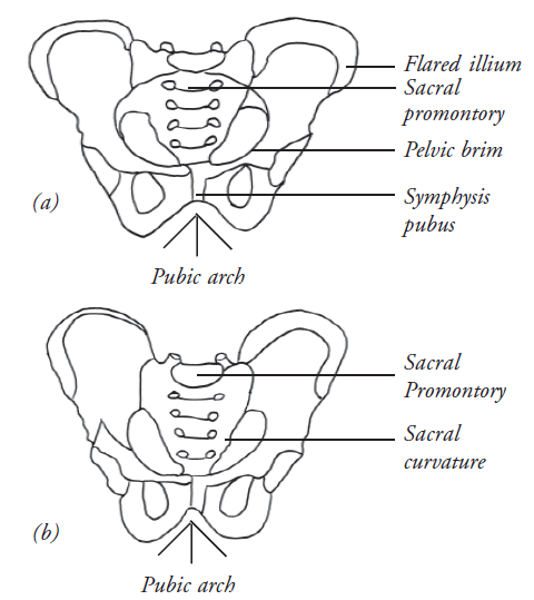
Pelvic Girdle and the lower limb
Pelvic girdle has 2 hip bones. The parts of the hip bone are: ilium, ischium, and pubis. The sacrum is formed by 5 vertebrae which are fused together.
Each lower limb has the following bones
| 1 femur/thigh | - the (longest strongest) bone of the upper leg |
| 1 tibia | - bone of the lower leg present in the medial side |
| 1 fibula | - bone of the lower leg present in the lateral side |
| 1 patella | - a triangular bone associated with knee joint |
| 7 tarsal bones | - bones of the ankle |
| 5 metatarsal bones | - bones of the dorsal part of the foot |
| 14 phalanges | - bones of the toes arranged similar to the fingers |
Joints
Joint is the site where two or more bones come together.
- Some joints are immovable joints, fibrous or fixed, for example : skull sutures
- Some joints are slightly movable or cartilaginous joints E.g. : symphysis pubis and the joints between the vertebrae
- Some joints are freely movable joints or synovial joints (wrist, elbow, knee, ankle)
3. Muscular system
This system is responsible for all types of movements of the body parts and its internal organs. Muscles cover the skeleton and are responsible for the shape of the body and make it firm. In this system we shall discuss
- Types of muscles
- Muscles of the face
- Muscles attached to the vertebral column
- Muscles of abdominal wall
- Muscles of trunk
- Muscles of upper limb
- Muscles of lower limb
Types of muscles
The body consists of two types of muscles
a. Voluntary muscles
b. Involuntary muscles
(a) Voluntary muscles are under our control. We can move them by our own will. They are connected to bones and move parts such as arms, legs and fingers.
(b) Involuntary muscles are not under our control. These muscles make many of the organs of the body work. The heart, stomach, urinary bladder, etc., are made up of involuntary muscles.
The proximal attachment of a muscle is called origin and its distal attachment is called either as fleshy fibers or as fibrous tendons. Depending on the arrangement of the muscle fibres with relation to the tendon, the muscles are divided into unipennate, bipennate and multipennate types. These muscles bring about movements in the joints.
Muscles of face joints
The muscles in the face producing movements of the lower jaw are called muscles of mastication. They are the masseter, temporalis and pterygoids. These muscles are attached to the mandible and are supplied by the mandibular nerve. These muscles also help in speech. Most of the muscles in the face and the scalp gain attachment to the skin and are responsible for the facial expressions. They are hence called the muscles of facial expression. Transverse wrinkles in the forehead are brought about by the occipito-frontalis muscle. The eyelids are closed by the muscle surrounding the orbit, the orbicularis oculi. The lips are moved by the orbicularis oris. The muscle that helps to blow air as in whistling is the buccinator. It is present in the region of the cheek. The angle of the mouth is elevated by the levator anguli oris and depressed by the depressor anguli oris. The dilation and compression of the nose are done by the nasalis. These muscles of facial expression are supplied by the facial nerve.
Muscles attached to vertebral column
The vertebral column is kept erect by the muscle sacrospinalis or erector spinae. The muscle is present in the posterior part of the vertebral column. It is supplied by the spinal nerves. The intercostal spaces are filled with muscles called the intercostals. They are supplied by the intercostals nerves. They help in respiration. However, the diaphragm is the most important muscles in inspiration. The muscle separates the structures in the thorax and abdomen. Contraction of the muscle results in the descent of the dome in the thoracic cavity. This leads to the expansion of the lungs and inspiration. The diaphragm is supplied by the phrenic nerve and the intercostals nerves.
Muscles of abdominal wall
The muscles of the anterior abdominal wall are arranged in three layers. They are the external oblique, internal oblique and the transverses abdominis muscles. On either side of the midline are the rectus abdominis muscles. These muscles help to bend the trunk forwards and laterally. The muscles are supplied by the intercostals nerves. The psoas major, quadratus lumborum and iliacus are the muscle of the posterior abdominal wall. They are supplied by the lumbar spinal nerves.
Muscles of trunk
The muscles trapezius, latissimus dorsi, pectoralis major and minor connect the upper limbs and the trunk. The trapezius is a muscle in the back. It extends between the occipital bone, cervical and thoracic vertebrae to the clavicle and scapula. The muscle produces movements of the clavicle and scapula. It is supplied by the spinal accessory and the cervical spinal nerves. The muscle latissimus dorsi is a broad sheet extending from the hipbone, lower ribs and thoracic vertebrae to the humerus. It is supplied by branches from the brachial plexus. The pectoral muscles are in front of the chest. The latissimus dorsi and the pectoral muscles help to adduct the upper limb.
Muscles of upper limb
The shoulder area is covered by a large multipennate muscle, the deltoid. It is an abductor of the shoulder joint. This muscle is selected to give intramuscular injections. The deltoid takes origin from the clavicle and scapula and is on the front of the arm. It has two heads of origin from the flexor of the elbow joint. Triceps brachii is a muscle in the back of the arm. It has three heads of origin from the scapula and the humerus. It is inserted into the ulna. It is supplied by the radial nerve and is a powerful extensor of the elbow joint.
The muscles in the front of the forearm produce flexion at the wrist joints. They are called the flexors of wrist. They are supplied by branches from the median and ulnar nerves. The muscles in the back of the forearm are the extensors of the wrist joint. They are supplied by branches from the radial nerve. The delicate movements of the fingers are brought about by the intrinsic muscles of the hand. These include the thenar, hypothenar, lumbricals and interossei. These muscles are supplied by branches from the median and ulnar nerves.
Muscles of lower limb
There are three large muscles in the gluteal region. They are gluteus maximus, gluteus medius and gluteus minimus.
This muscle mass is selected to give intramuscular injections. The gluteus maximus produces extension of the hip joint and the gluteus medius and minimus produce abduction of the hip joint. They are supplied by branches from the sciatic plexus. The muscle in the front of the thigh is the quadriceps femoris. It has four heads into the patella and extends as the ligamentum patellae to the tibial tuberosity. The muscle is supplied by the femoral nerve and it helps in the extension of the knee joint. The hamstring muscles are present in the back of the thigh. They extend from the hip bone to the bones in the leg. They are supplied by the sciatic nerve. Adductor longus and adductor magnus are situated in the medial aspect of the thigh. They are supplied by the obturator nerve and produce adduction of the hip joint. The muscles in the anterior part of the leg are extensors. They produce dorsiflexion of the ankle joint. The flexors are supplied by anterior tibial nerve and the extensors are supplied by the posterior tibial nerve. The movements of the foot are brought about by the intrinsic muscles of the foot. They are the muscles of great toe, muscles of little toe, lumbricals and interossei. They are supplied by planter nerves, branches from the posterior tibial nerve.
4. The cardiovascular system
This system deals with heart, blood and blood vessels. They function effectively in nourishing all other organ systems of the body. Here we discuss
- Structure and function of heart
- Circulation of the body
- Constituents of blood and their functions
- Functions of cardiovascular system
The Heart
The heart is the pumping organ which maintains blood circulation throughout the body. Heart is a conical, hollow, muscular organ, situated between the two lungs. The heart is about the size of the owner’s fist (Fig. 3.7).

Layers of the Heart
The heart is composed of three layers:
- Pericardium
- Myocardium
- Endocardium
Interior of the Heart
The heart is a four chambered organ. It has two atrium and two ventricles. Atria and ventricles are divided into right and left side by a continuous partition called the inter atrial septum and inter ventricular septum. Atrioventricular valves separate atrium and ventricle. These valves allow blood to flow only in one direction, i.e. from the atrium to the ventricle. Semilunar valves are present at the opening of the aorta and pulmonary artery.
Flow of blood through the Heart
The superior vena cava and the inferior vena cava empty the deoxygenated blood into the right atrium. This blood passes into the right ventricle. From there it is pumped into the pulmonary arteries (the only artery which carries de-oxygenated blood). Pulmonary arteries carry venous blood to the lungs for purification. In the lungs carbon dioxide is excreted and oxygen is absorbed. The oxygenated blood is carried to the left atrium by two pulmonary veins from each lung. It then passes through the left atrioventricular valve into the left ventricle, and from there it is pumped into the aorta. Through aorta the blood is pumped into various parts of the body.
Blood
The human body has 5-5 ½ litres of blood. It consists of plasma and blood cells.
Plasma is a straw coloured transparent fluid. It contains water, plasma proteins, minerals, nutrients from digested foods, hormones, clotting factors, antibodies, gases, and waste materials like urea.
Arteries
Blood vessels that carry oxygenated blood from the heart are called arteries (Fig. 3.8). The artery branches many times to smaller arteries and finally end as arterioles. The major blood vessels of the body are: aorta, carotid, brachial, radial and femoral.
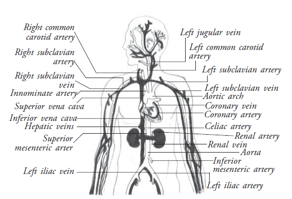
Veins
The veins are the blood vessels that return the impure blood to the heart.
Capillaries
The smallest arterioles breakup into a number of minute vessels called capillaries. They form a network which enables the exchange of substances, i.e. gases and nutrients.
Functions of the circulatory system
- The system takes oxygen from the lungs to other parts of the body. It also takes carbon dioxide from different parts to the lungs.
- The nutrients of the digested food are taken from the intestines to the liver.
- The digested food is converted to glucose in the liver. From the liver it reaches all other organs through the blood. The energy from the liver is taken to all parts of the body.
- Waste products are taken to the kidneys.
- Hormones from the endocrine glands are taken to their place of action.
- It keeps the temperature of the body constant.
- It keeps the water content in the body constant.
- The leucocytes in the blood fight against diseases.
- The platelets in the blood play a vital role in the coagulation of blood when vessels are damaged.
Medical terminologies
| Systole | : Contraction of both atrium and ventricles. |
| Diastole | : Relaxation of both atrium and ventricles. |
| ECG | : Electrocardiogram (ECG ) is done to detect the electrical activity within the heart, which can be obtained on a print out. |
| Blood pressure | : It is defined as the force or pressure that the blood exerts on the walls of the blood vessels. The normal diastolic pressure is 120 mm of mercury. The normal diastolic pressure is 80 mm of mercury. |
| Pulse | : Pulse is the wave of distension felt in an artery wall, when blood is pumped out of the heart. (Normal 72-80 / minute) |
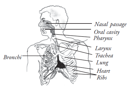
5. Respiratory system
All cells in the body need oxygen to carry out their activities. The respiratory system (Fig. 3.9) deals with breathing. When we breathe, the body takes in the oxygen that it needs and removes the carbon dioxide that it doesn’t need . In this system we shall discuss
- Respiratory pathway
- Structure of lungs
- Mechanism of respiration
Respiratory pathway
Anatomy of the respiratory system
Nasal cavity
Air enters the nasal cavity through two nostrils. The nose preconditions the air that enters the lungs, by cooling it or warming it and by removing dust etc.
Sinuses
Sinuses are hollow spaces in the bones of the head. Small openings connect them to the nose. The functions they serve include
1. Helping to regulate the temperature and humidity of air breathed in.
2. To lighten the bone structure of the head and to give resonance to the voice.
Pharynx
Commonly referred to as the throat. The throat is the common pathway for air and food. The air passes through the larynx into the trachea while food passes into the esophagus.
Larynx
This is the voice box and contains vocal cords in its interior. The opening of the larynx into the trachea is a narrow slit called the glottis, through which air passes into the trachea. Glottis is covered by epiglottis that keeps food out of the respiratory tract during swallowing.
Trachea / wind pipe
This is a tubular structure. It has 'C' shaped cartilage rings. They maintain the tubular structure of the trachea. The windpipe divides into the two main bronchial tubes that enter into each lung. They subdivide further for each lobe of the lungs. In the lobe they again subdivide for each lobule.
Structure of lungs
There is a pair of lungs enclosed in the thoracic cage. A double-layered membrane called pleura covers the lungs. There is a fluid between the two membranes, called pleural fluid. It lubricates and minimizes the friction between the two membrane. The right lung is divided into three lobes or sections. Each lobe is like a balloon filled with sponge-like tissue. The left lung is divided into two lobes. The space between the two lungs is called mediastinum, where the heart is located.
The Alveoli
The alveoli are very small air sacs. They are the destination of the air we breath in.
Breathing
Breathing is the process by which oxygen in the air is taken into the lungs and brought into close contact with the blood. Blood absorbs it and carries it to all parts of the body. At the same time the blood gives up waste matter (carbon dioxide), which is taken out of the lungs when air is breathed out.
The mechanism of respiration
First the body breathes in the air which is sucked through the nose or mouth and down through the trachea, bronchus, and into the lungs. Inside the lung the air reaches the alveoli. In the walls of alveoli, tiny blood vessels allow exchange of O2 & CO2 in capillaries.
6. Nervous system
The nervous system detects and responds to the changes inside and outside the body. For descriptive purposes, the nervous system is divided as follows.
Central nervous system, autonomous nervous system and peripheral nervous system.
Central nervous system
The central nervous system consists of the brain and the spinal cord. The dura matter, the arachnoid matter and the pia matter are the three layers of membranes covering the brain and the spinal cord. Inside the brain there are cavities called ventricles, which contains cerebral spinal fluid (CSF). It acts as a cushion and shock absorber. It also protects the brain and spinal cord.
Brain
The brain (Fig. 3.10) constitutes about one fifth of the body weight. It lies within the cranial cavity.
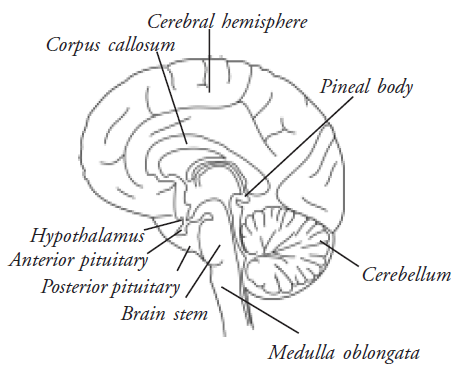
The parts of the brain are
- Cerebrum
- Midbrain
- Pons
- Medulla oblongata
- Cerebellum
Cerebrum
- Cerebrum
- Midbrain
- Pons
- Medulla oblongata
- Cerebellum
Cerebrum
Cerebrum is divided into four lobes
Frontal - Parietal
Temporal - Occipital
Functions of the cerebrum
- Memory
- Intelligence
- Thinking
- Reasoning
- Moral sense
- Learning
In the grey matter there are two areas called the hypothalamus and thalamus. They control body temperature, hunger and thirst, emotional reactions, sexual behaviour (including mating and child rearing), biological clocks (e.g. sleeping and waking cycles), secretion of some hormones.
Midbrain and pons
These two act as relay stations between the brain and the spinal cord.
Medulla oblongata
Medulla oblongata has the vital centers in its deeper structure.
The spinal nerves
These nerves leave the spinal cord through inter vertebral foramina. They are named and grouped according to the vertebrae to which they are associated.
- 8 Cervical
- 12 Thoracic
- 5 Lumbar
- 5 Sacral
- 1 Coccygeal
The lumbar, sacral and coccygeal nerves leave the spinal cord near its termination at the first lumbar vertebra and extend downwards inside the vertebral canal. This resembles a horse's tail and so is called " Cauda equina ". All spinal nerves have an anterior nerve root (motor) and posterior nerve root (sensory). So they are mixed nerves.
The cranial nerves
There are 12 pairs of cranial nerves in our body. Some are sensory, some are motor and some are mixed nerves. They are:
- Olfactory
- Optic
- Oculomotor
- Trochlear
- Trigeminal
- Abducent
- Facial
- Vestibulocochlear (auditory)
- Glossopharyngeal
- Vagus
- Spinal accessory
- Hypoglossal
The autonomous nervous system
The autonomous nervous system controls the functions of the body which are carried out automatically. For example
- Rate and force of the heart beat
- Secretion of the glands
- Vasoconstriction or vasodilation
- Change in the size of the pupils of the eye
The autonomic nervous system is divided into two parts.
- Sympathetic nervous system
- Parasympathetic nervous system
Specialised centers of the brain are
| Cardiac center | - Controls the rate and force of cardiac contraction. |
| Respiratory center | - Controls the rate and depth of the respiration. |
| Vasomotor center | - Controls the diameter of the blood vessels. |
| Reflex centers | - Eliminate irritating substances by reflex actions of vomiting, sneezing and coughing. |
Cerebellum
The cerebellum is situated behind the pons and it plays an important role in maintaining posture and balance.
Spinal cord
The brain is connected to the spinal cord. In the spinal cord, the gray matter is found in the center and white matter surrounds it. The opposite arrangement (white matter in the center and gray matter surrounding it) is found in cortex of the brain. All the messages that travel from the brain pass through the spinal cord and then to all parts of the body through the nerves. Messages from different parts of the body also pass through the spinal cord on their way to the brain. The spinal cord is protected by the vertebral column.
The peripheral nervous system
The peripheral nervous system consists of 31 pairs of spinal nerves.
Sense organs
Human beings are gifted with sense organs, which are means of communication to the external world. Human body consists of five sense organs which have special senses.
- Ear
- Eye
- Nose
- Tongue
- Skin
Ear
Anatomy of ear
There is a pair of ears in the human body. The ear (Fig. 3.11) is the organ used for sensing sounds, i.e., the organ used for hearing. The ear can be divided into three parts:
- External ear
- Middle ear
- Inner ear
Structure of ear
External ear
The external ear or the pinna is used to receive the sound waves.
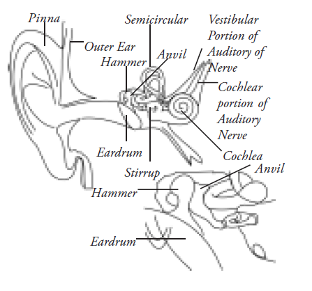
Middle ear
The middle ear consists of the three smallest bones of our body, They are stapes, incus and malleus. The malleus is attached to the tympanic membrane. These bones transmit sound waves from the external auditory canal to the inner ear.
Inner ear
The inner ear consists of three semicircular canals, utricle, saccule, endolymphatic duct and the cochlea. The auditory nerve from the cochlea transmits the sound impulses to the brain, which identifies the sound.
Physiology of ear
Sound waves are conducted through air in the external ear. They are conducted through the bones in the middle ear. From here they pass through fluid in the inner ear. The stages involved in the transmission of sound waves are as follows:
- Sound vibrations enters the external auditory meatus / ear canal, tympanic membrane (ear drum).
- The vibrating membrane transmits the sound to the handle of malleus (hammer) which is attached to the eardrum at about its midpoint.
- Bone conduction of sound waves proceeds through the three tiny connected ear bones from malleus to incus to stapes.
- Sound is then transmitted from the footplate of the stapes through the oval window of the vestibule of the inner ear. Pressure is thus exerted by the foot plate inward into the perilymph (fluid) in the scala vestibuli of the cochlea.
- The sound ripple passes through this fluid and finally it is expanded against the round window.
- The message is gathered from here by the cochlear nerve and reaches the brain.
Nose
Man can sense smell with the help of the nose. The nose consists of 2 nostrils. The nostrils are lined with mucus membrane. The nose is bounded by cartilage and bone. The nose is lined by olfactory epithelium, which consists of supporting cells, basal cells and the olfactory receptor cells. These are highly specialised cells from which minute fibers pass to join the olfactory bulb. The olfactory bulb is the slightly enlarged portion of the olfactory tract which lies above the ethmoid bone.
The sensation of smell is stimulated by gases inhaled. The act of sniffing causes the nostril to dilate and the direction of the anterior part of the nasal respiratory chamber is altered, so that the stream of inspired air is directed towards the upper olfactory area of the cavity. Odours can be finely perceived in very minute quantities and can be finely discriminated. The sense of smell is lessened if the nasal mucous membrane is very dry, wet, or swollen as in a cold in the head. Smells are described as pleasant or unpleasant.
There is an inter-relationship between smell and taste sensations; people complain of impaired taste when they have nasal congestion.
Tongue (sense of taste)
The tongue is the organ used in identifying taste. It also assists in speech. It mixes saliva with food, it aids in chewing and swallowing. The tongue is a solid mass (Fig. 3.12) of muscle fibers.
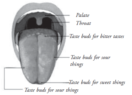
Structure of tongue
The dorsal surface of the tongue is covered with numerous minute projections or papillae, which vary in shape. The largest are the vallate present in the root of the tongue. The most numerous are the thread like filliform papillae, and the fungiform papillae (mushroom shaped) which are present at the tip of the tongue.
The root of the tongue is supplied with lymphoid nodules called the lingual tonsil. The receptor of taste, the taste buds are distributed on the dorsal surface of the tongue. They serve to distinguish the four basic tastes – salt, sweet, sour and bitter.
When the food is chewed, it gets mixed with saliva. The juice penetrates the taste buds, and enables them to identify the taste.
The musculature of the tongue is supplied by the hypoglossal nerve (12th cranial nerve).
Skin
Skin is the outer most layer of the human body. It protects the inner organs from various external factors. The external appearance of the skin is deceptively homogeneous and simple. It consists of two layers.
The outer layer - epidermis / cuticle
The outer layer, the epidermis is made up of five or six layers of cells. They consist of a cornified layer of dead cells. Cells are shed from this layer and replaced by cells that are produced in the basal layer of the epidermis (Fig. 3.13).
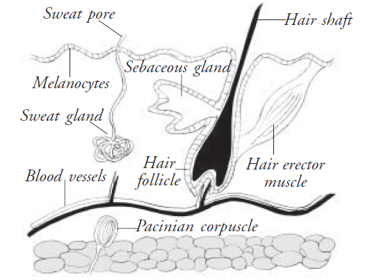
The inner layer - dermis / corium
The dermis is composed of dense, white connective tissue. It is supportive in nature. This contains blood vessels and sensory nerve endings for pressure, touch, pain and temperature. Sweat and oil (sebaceous) glands are present in the dermis. The hair follicles arise from the dermis. The sebaceous glands are located near the hair follicle. The sweat glands open out through sweat pores, which are numerous in the epidermis.
Functions
- Skin is an organ of temperature regulation.
- Skin is a vascular organ which plays an extremely important role in temperature regulation.
- The blood vessels to the skin are organized in a way that blood may be shunted from the arteries to veins when the body needs to preserve the heat and vice versa. When a person does prolonged muscular exercise, the blood flow to the skin will increase, thereby releasing heat.
- The skin also regulates temperature by sweating.
- When the body sweats a lot, it releases heat thereby reducing the body temperature.
Melanin
The skin colouration is due to the pigment called melanin. If the melanin pigment is more, the skin appears dark.
Skin is an organ of the sense of touch, heat, cold, and pain resulting from the stimulation of the nerve endings in the skin. The sense varies with the type of nerve ending stimulated.
Skin is an organ of storage
The skin and the underlying tissue act as storage for water. The adipose tissue beneath the skin is one of the principal fat depots of the body.
7. Excretory system
The waste products from the body are eliminated through the excretory system. In this system we discuss
- The excretory substances
- The excretory organs
- The structure of kidney
- Formation of urine
- Functions of kidneys
The excretory substances
The excretory system is responsible for the elimination of wastes produced by homeostasis. Metabolism of the various chemical substances produces different waste products. They are:
- Carbondioxide
- Urea and nitrogenous waste
- Salts
- Excess water
- Undigested food
- Toxic substances
- Pathogenic organisms
Excretory organs
There are
- Sweat Glands
- Liver
- Lungs
- Kidneys
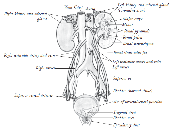
Kidneys
The principal excretory organs are kidneys. (Fig. 3.14) Every human has two kidneys, which remove urea from the blood. The kidneys are bean-shaped and dark red in color. They are situated inside the abdomen on either side of the vertebral column. The kidney is composed of two layers.
Outer layer - the cortex
Inner core - the medulla.
Formation of urine
Each kidney is composed of a number of functional units called nephrons. There are 1 million nephrons in each kidney. Nephrons are the functional units of the kidneys. The tops of the nephrons are in the cortex, while their long tubule portions are in the medulla.
The nephron consists of the glomerulus, Bowman’s capsule, proximal tubule, loop of Henle, the distal tubule and the collecting ducts.
The structure and the functions of the parts of the kidneys are given below
| Glomerulus | : This is the tuft of capillaries through which the blood comes to nephron for filtration |
| Bowman 's capsule | : This filters the blood, which comes through glomerulus |
| Proximal tubule | : Here water and some " good " molecules are absorbed back into the body, while a few other, unwanted molecules / ions are added to the urine. |
| Loop of Henle | : Here more water is removed (back into the bloodstream) Some NaCl (salt) is removed from the filtrate at this point to adjust the amount of fluid which surrounds the tubule. Capillaries wind around and exchange materials with the tubule. |
| Distal tubule | : Here more water and some " good " solutes are removed from the urine, while some more unwanted molecules are put in. |
| Collecting duct | : Gathers urine from several nephrons. As the collecting duct goes back through the medulla, more water is removed from the urine. |
| Renal Pelvis | : The collecting ducts eventually end up at the renal pelvis, which collects the urine from all of them. |
| The ureter | : From the renal pelvis of each kidney the ureter conveys the urine to the urinary bladder. |
| Urinary bladder | : This is a pear shaped bag where the urine is temporarily stored. |
| Urethra | : This conveys urine from the bladder to outside the body. |
Functions of the kidneys
- Excretes urea formed during protein metabolism.
- Excretes toxic substances.
- Regulates the loss of excess water from the body.
- Maintains the pH.
- Secretes a hormone called erythropoietin responsible for development of RBCs.
- Converts inactive vitamin D into active vitamin D.
When waste products are removed properly, the body is healthy. When the kidneys are infected, the quantity of the urea in blood will increase, endangering life.
Glucose is not normally excreted. But in the case of diabetic patients, it is excreted in urine. When diseases affect a person, the qualities of the urine change.
8. Digestive system
Food provides us with fuel to live, energy to work and play, and the raw materials to build new cells. All the different varieties of food we eat are broken down by our digestive system and transported to every part of our body by our circulatory system. In digestive system we discuss .
- Organs of the digestive system
- Functions of alimentary tract
- Liver
- Large intestine
- Rectum and anus
Organs of the digestive system
The Digestive system includes the alimentary canal and the digestive glands. The alimentary tract has the following parts
- Mouth
- Pharynx
- Esophagus
- Stomach
- Small intestine
- Duodenum
- Jejunum
- Ileum
- Large Intestine
- Appendix
- Cecum
- Colon
- Rectum
- Anus
The accessory glands
- Salivary glands of the mouth
- Parotid
- Sub mandibular
- Sublingual
- Liver
- Pancreas
- Biliary tract
The wall of the alimentary canal (except for the mouth and the pharynx) is made up of four layers from outer to inner. They are
- Peritoneum
- The muscular layer
- The submucosa
- The mucosa
The alimentary tract
- Teeth grind the food; the tongue helps to chew food and mix it with saliva to make a soft pulp that is easy to swallow.
- The stomach secretes the following enzymes:-
- Hydrochloric acid
- Pepsinogen
- Rennin
- Gastric amylase
- The secretions of small intestine is called Succus Entericus. The small intestine is divided into duodenum, jejunum and ileum. The bile and the pancreatic juice enters the duodenum. This has lipase and amylase which digest the fat and carbohydrates respectively. The small intestine secretes intestinal juice which digests the food into nutrients that are small enough to pass through the lining of the small intestine and into the blood. They are carried away to the liver and other parts of the body to be processed, stored and distributed.
Liver
Blood from the intestines flows to the liver, carrying nutrients, vitamins and minerals, and products from digestion. The liver is like a food processing factory with more than 200 different jobs.
- It stores nutrients
- It changes absorbed nutrients from one form to another (Glucose into glycogen).
- It releases them into the blood according to the activities and needs of the body.
Large intestine
The large intestines is made up of the colon, cecum and appendix. Any useful substance in the leftovers, such as spare water and body minerals, are absorbed through the walls of the large intestine, back into the blood. The remains are formed into brown, semisolid faeces, ready to be removed from the body.
Rectum and anus
The end of the large intestine and the next part of the tract, the rectum, stores the faeces. These are finally squeezed through the anus out of the body.
9. Endocrine system
The endocrine glands or ductless glands secrete certain chemical substances called hormones. They guide and control the various metabolic activities, and the growth and differentiation of various systems. These glands do not have any ducts. As secretory glands they directly pass their secretion into the blood stream. In this system we discuss.
- General principles of hormones
- The different endocrine glands and their functions
- Gonads and reproductive system
General principles of hormones
- Hormones are secreted in response to specific stimuli.
- Hormones can be secreted independently of one another.
- Hormones are present in minute quantities in the blood.
- Hormones are believed to be catalytic in their effects.
- Hormones have a high degree of target specificity.
- All hormones are proteins in nature.
- Hormones can be extracted artificially.
- Hormones can be synthesized, isolated and purified.
Endocrine glands
- Pituitary gland
- Thyroid gland
- Parathyroid gland
- Islets of Langerhans (Pancreas)
- Adrenal glands
- Gonads
- Thymus
Pituitary gland [Hypophysis]
This is the chief center of the endocrine system. It forms an important link between the nervous and endocrine systems. It controls other endocrine glands. It is a small nut like structure situated at the base of the brain. It is divisible into two distinct portions – the anterior lobe (adenohypophysis) and the posterior lobe (neurohypophysis).
Hormones secreted in adenohypophysis
- Growth hormone: This hormone is responsible for growth of cells and tissues. Deficiency of this hormone results in Dwarfism. Excessive secretion results in Gigantism (Acromegaly).
- Thyroid stimulating hormone: It stimulates adrenal glands to synthesise and release its hormones.
- Adrenocorticotrophic hormone (ACTH): It stimulates adrenal glands to synthesise and release its hormones.
- Follicle stimulating hormone: It stimulates the growth of Graffian Follicle in the ovary in females and promotes the formation of sperm in the testes of males.
- Luteinizing hormone: It plays an important role in causing ovulation in females and also in development of corpus luteum. In males it stimulates the interstitial cells of the testes to secrete testosterone.
- Prolactin: It is associated with lactation in females.
Antidiuretic hormone (Vasopressin) :
(ADH) It causes the kidneys to retain water, thus increasing the water content of the body. It plays an important role in osmoregulation / water balance of the body.
Oxytocin:
It causes rhythmic contraction of the uterus at the time of childbirth for the expulsion of the fetus and the placenta. It causes the ejection of milk from the lactating mammary glands.
Thyroid gland
The gland consists of a pair of lobes. They lie one on either side of the larynx in the neck region, they are connected by a median structure called isthmus. This gland secretes a hormone called thyroxine. Iodine is essential for the production of thyroxine. Iodine deficiency leads to goiter.
Functions of thyroxine
- Stimulates the normal growth and development
- Controls the rate of cellular oxidation and basic metabolic activities.
Disorders of thyroid gland are
- Deficiency (hypothyroidism) in children leads to cretinism. In adults leads to myxedema.
- Excessive secretion results in hyperthyroidism.
Parathyroid glands
They are four small glands placed posteriorly 2 on each side of the thyroid. They secrete (1) Parathormone and (2) Calcitonin.
Functions
They control the metabolism and blood levels of calcium and phosphate.
Disorders
Hypoparathyroidism - Tetany due to fall of blood calcium level
Excessive secretion - Hyperparathyroidism
Islets of Langerhans
The islets of Langerhans are groups of epithelial cells found lying between the portions of pancreas. They contain alpha, beta and delta cells. Alpha cells produce Glucagon. Beta cells produce Insulin. Delta cells produce Somatostatin
Role of insulin
It increases the withdrawal of glucose from blood in 3 ways, or reduces the glucose level in the blood.
- Conversion of glucose into glycogen
- Oxidation of glucose in the tissues.
- Conversion of glucose into fat.
Deficiency causes hyperglycemia (increase of glucose in the blood). This disease is known as diabetes mellitus. Excess secretion lowers the blood sugar leading to hypoglycemia.
Role of glucagon
Its effects on blood glucose level are generally opposite to the effects of insulin. It increases the release of glucose from glycogen. A proper balance between insulin and glucagon production is therefore necessary to maintain proper blood glucose levels.
Adrenal glands
The two adrenal glands lie on the upper pole of the kidneys. They are also called supra renal glands.
Hormones secreted
Adrenal cortex
Aldosterone - Promotes reabsorption of sodiumions from the renal glomerular filtrate
Cortisone - Production of glucose from non- carbohydrate sources like fats and amino acids. It also acts as an anti-inflammatory agent
Adrenal medulla
- It secretes adrenaline (epinephrine), non adrenaline (non epinephrine)
- Gives instant energy,
- Stimulates constriction of blood vessels
- Increases the heart rate.
- Relaxation of the smooth muscles
- Causes goose flesh.
- Breakdown of glycogen to glucose
Gonads
They produce the sex hormones in the ovaries of females and in the testes of males.
10. Reproductive system
Reproduction is the only means by which the continuity of life is maintained. Human beings reproduce sexually. Male and female reproductive organs carry out the process of sexual reproduction which begins with fertilization of ovum by the sperm.
Female reproductive system
This consists of external genitalia - a pair of ovaries, a pair of fallopian tubes, uterus and a vagina.
Ovaries
One pair of ovaries is located in the abdominal cavity. The ova begin to develop inside the ovaries. The ovaries of the sexually matured female contain ova at various stages of development. The final stage is a structure called the Graafian follicle, which consists of an ovum. The graffian follicle ruptures and the ovum is released.
Fallopian tubes
The terminal part of the fallopian tube is a funnel shaped structure, lying close to the ovary. It receives the ova from ovary and conveys into the uterus.
Uterus
Uterus is a muscular organ situated in the pelvic cavity.
The uterus consists of three layers
Perimetrium - Outer layer
Myometrium - Middle layer
Endometrium - Inner layer
The main function of the uterus is to prepare the endometrium for reception of fertilized ovum. The fertilized ovum is embedded in the uterine cavity where it develops and is nourished to full term. If the ovum is unfertilized, the endometrium is shed off as menses.
Vagina
This is the muscular tube lined with mucous membrane connecting the cervix at the upper end and external genitalia at the lower end. It receives sperms and serves as birth canal.
Male reproductive system
This consists of a pair of testes, seminal tracts and related glands.
Testes
There is one pair of testes inside the scrotum. The scrotum is a skin bag having two separate compartments, one for each testes. The testes form the male gametes known as spermatozoa. Each testes consists of a large number of tubules that secrete the hormone testosterone.
Epididymis
The tubules in the testes unite to form a long coiled structure called epididymis. It lies on the outside of each testis and is attached to it. This is the main storehouse of sperm. The sperm attain their motility in the epididymis.
Vas deferens
The epididymis differentiates into a muscular tube called the vas deferens. It ascends and reaches via abdominal cavity into the pelvis, opening into the urethra.
Seminal vesicles
These are paired tubular glands situated behind the neck of the urinary bladder. Each seminal vesicle’s duct joins with the vas deferens of that side.
Urethra
After leaving the urinary bladder, urethra passes through the prostate gland, where it is known as the prostatic urethra. Both the vas deferences and the urethra join here.
Prostate gland
This is a gland composed of fibromuscular and glandular tissue. It is located at the lower end of the urinary bladder, it surrounds the first part of the urethra.
Penis
It is composed of spongy erectile tissue through which the urethra (penile urethra) traverse and opens to the exterior.
Practical applications
Surface Anatomy
The important surface markings related to human anatomy
- Thyroid gland is situated in front of the neck just below the thyroid cartilage.
- The heart lies in the chest. Surface marking: Two finger breadth from the lower most part behind the chest bone, extending to the left between 2nd and 5th ribs. The apical impulse can be felt in the 5th left inter coastal space below the nipple.
- To start intravenous lines, antecubital vein is commonly used. This is situated on the anterior aspect of the elbow.
The Pulse can be palpated at
Carotid Artery
The carotid arteries can be recognized in the lateral part of the neck just lateral to the larynx (voice box).
Brachial artery
The brachial artery is situated in the anterior part of the arm and can be palpated in the anterior part of the elbow.
Radial artery
The radial artery can be palpated in the forearm. Surface marking: just above the wrist on the lateral part of forearm.
Femoral artery
Femoral artery can be palpated on the upper part of the thigh
Dorsalis pedis artery
Dorsal pedis artery is situated on the anterior aspect of the foot.
Other important practical points
- Insulin is produced in the pancreas. Insulin, along with other hormones of glucagon, corticosteroids, adrenaline are important in maintaining proper blood glucose levels in the body. Lack of insulin causes diabetes.
- Excess or lack of thyroid hormone causes hyper/hypothyroidism. Hyperthyroidism can cause manifestation in the eye (proptosis)
Summary
This unit has covered each organ in detail. All the systems of our body are interdependent and hence any dysfunction of an organ can cause serious effect to our eye. Therefore, it is very important to take the patient's history in detail when they come with any ocular ailment.
Key points to remember
- The organs of a living organism carry out activities that maintain life. Different organs form an organ system and carries out a particular function.
- The major systems of the human body are skeletal, muscular, respiratory, circulatory, digestive, nervous, excretory, sense organs, endocrine and reproductive.
- The respiratory organs are the trachea, bronchi and lungs. Respiration involves the exchange of oxygen and carbon dioxide and the oxidation of food in each cell to release energy.
- The heart, blood vessels and blood make up the circulatory system. These transport nutrients, oxygen, hormones and various other substances throughout the body.
- The kidneys, lungs, skin and large intestine are the excretory organs of the body.
- Digestion means converting the complex food we eat into a simpler form that is easily absorbed by the body.
- Food goes through numerous digestive organs like the mouth, esophagus, stomach, small intestine and large intestine. The liver and pancreas secrete certain juices that help in digestion.
- The brain, spinal cord and nerves form the nervous system. It controls and coordinates all the activities of our body and the functions of other organ systems.
- Nerves carry messages throughout our body. There are 31 pairs of spinal nerves and 12 pairs of cranial nerves.
- Some nerves carry messages from the brain to the other parts of the body. Others carry messages from different parts of the body to the brain.
- The 206 bones of the skeletal system give shape and support to the body. It also protects organs like the heart, brain and lungs.
- The muscles of the muscular system cause movements of various parts of the body and internal organs.
- The organs of the reproductive system are the testes in males and ovaries in females. They produce reproductive or sex cells.
- Testes produces sperm and the ovaries produce ovum.
Student exercise
A. Tick the most appropriate answer
1. Sperms are produced in
| a. Ovaries | b. Testes |
| c. Eggs | d. Urethra |
2. The skeletal system consists of
| a. Muscles | b. Bones |
| c. Muscles and bones | d. Brain |
3. The food does not pass through these glands but they help in digestion
| a. Liver and pancreas | b. Small intestine and pancreas |
| c. Liver and heart | d. Large intestine and stomach |
4. In clotting the blood _______ plays an important role
| a. Red blood cells | b. White blood cells |
| c. Thrombocytes | d. Arteries |
5. It act as the relay station between the brain and the spinal cord
| a. Mid brain and | b. Cerebellum & pons |
| c. Frontal and parietal | d. Occipital and temporal |
B. Fill in the blanks
- The artery which carries deoxygenated blood is ________________.
- ______________ part of the brain is responsible for memory.
- Insulin is secreted by _______________.
- The covering of the lungs is called ______________.
- The basic functional unit of kidney are __________________.
- The male reproductive system consists of _________ that produce sperms
- The nervous system consists of the brain,______ and _________.
- The conversion of complex food into simpler absorbable form is called _______.
C. True or False
- The excretory system carries oxygen to all the parts of the body.
- Femur and patella are responsible for the bone formation.
- In the circulatory system, the brain, spinal cord and nerves coordinate all the functions of the body.
- Tissues are grouped together to form systems.
- Muscular system is responsible for all types of movements of the body parts and its internal organs.
- Pharynx is commonly referred to as voice box and larynx is referred to as throat.
- Pituitary gland is the chief center of the endocrine system.
D. Match the following
- Small intestine - exchange of gases
- Artery - shape to the body
- Bones - situated in the pelvic cavity
- Urethra - transporting urine to the urinary bladder
- Respiratory system - digestive system
- Ureter - opening of the urinary bladder
- Muscular system - carry oxygenated blood
- Uterus - skeletal system
E. Find the odd one out. Give reasons
- Oesophagus, trachea, small intestine, anus
- Nostrils, lungs, bronchi, nerves
- Brain, spinal cord, arteries, nerves
- Kidney, stomach, ureter, urethra
- Veins, capillaries, arteries, canines
F. Rearrange the organs to show
- The path of the food in the body Small intestine, stomach, mouth, large intestine, oesophagus
- The path of the oxygen rich air in the body Lungs, nose, trachea, bronchi
- The path of blood from the heart capillaries, heart, veins, arteries
G. Give the values of
- Blood volume
- Pulse
- Systolic Pressure
- Diastolic pressure
- Number of bones
- Spinal nerves
- Cranial nerves
- Vertebrae
- Nephrons in both kidney
- Number of lobes in right lung
- Number of lobes in left lung
H.Explain the following
- Power house of cell
- Floating ribs
- Voice box
- Throat
- Wind pipe
- Brain stem
- Succus entericus
- Graffian follicle
- Axis
- Atlas
I. Differentiate the following
- Ureter and Urethra
- Neuron and Nephron
- Glucagon and Insulin
J. State the opposites of
- Adduction
- Depression
- Spermatogenesis
- Sympathetic
- Osteoclast
- Bicuspid valve
K. Answer the following
- Describe the functions of cardio vascular system
- Identify the excretory organs and the waste eliminated by them
- Describe the path of food through the digestive system
- Describe the functions of the nervous system.
- What are the basic differences between voluntary and involuntary muscles?