Anatomy and Physiology of the Eye
Introduction
The eye is the most sensitive part of our body. It views the world. It is also exposed to different light of the object. The human body has a pair of eyeballs. The eyeballs are well protected in bony cavities called orbits which are situated on either side of the nose.Externally the eye is protected by the eyebrow, eyelashes and eyelids.
Development of the eye
The embryonal and fetal development of the human eye includes a series of sequential events starting with the fertilization of the ovum and ending with birth of a normal baby. Three main periods can be distinguished in the development of the eye.
The first period called embryogenesis is characterized by appearance and migration of the neural crest cells and by the formation of the primary brain vesicles known as optic vesicle and ends at the end of the 3rd week. The second period called organogenesis extends from the 4th week till the end of the 8th week. It includes the closure of the neural canal interiorly with the subsequent evagination of its lateral wall into optic vesicles, the invagination of the lower nasal wall of the optic vesicle causing the formation of the optic cup, and the development of the lens plate, retinal disk and embryonic fissure. The embryonic fissure extends into the optic stalk which connects the cavity of the optic vesicle with the cavity of the neural canal; the hyaloid artery penetrates into the optic cup through the embryonic fissure. The primary vitreous forms and the surface ectoderm overlying the lens vesicle differentiates into a primitive corneal epithelium. The third period involves the differentiation of each of the primitive organs into a fully or partially active organ and is called differentiation. The facial and orbital structures also develop at this stage.
Schedule of eye development
| 3.5 wks. | Optic vesicle appears, set at 180° to each other |
| 4th wk. | Optic cup involutes and lens placode/pit forms. |
| 5th wk. | Lens vesicle separates from epidermis; choroid fissure present in optic cup; vitreous appears. |
| 6th wk. | Retina differentiated into neural and pigment layers; lens thickens; eyes rotate to 160°. |
| 7th wk. | Choroid fissure closes; lens cavity obliterated; lids begin to form; axons enter optic stalk. |
| 10th wk. | Eyelids fuse; ciliary body and iris forming. |
| 12th wk. | Layers of retina are organising. |
| 21st wk. | Retinal layering complete, responsive to light; eyelids open. |
The orbit
The orbit is a pyramidal bony cavity containing the eyeball, extraocular muscles, fat, blood vessels and important nerves. It is formed by a roof, floor, medial wall, lateral wall, a base and an apex. The bony orbit is made up of 7 bones - frontal, sphenoid, zygomatic, maxillary, palatine, ethmoid and lacrimal. Five important cranial nerves (2,3,4, first division of 5 and 6) pass through the apex of the orbit.
External structure of the eye
Eyebrows
The eyebrows are formed by several rows of hair. They separate the upper lid from the forehead. Besides protection, they contribute to facial expression by their movement
Eye lashes
Eye lashes arise from follicles arranged in irregular rows. There are 5-6 rows in the upper lid and 3-4 rows in the lower lid.
Canthi
The site of the union of the upper and lower eyelids near the nose is known as medial canthus and the site of union towards the ear is known as lateral canthus.
Caruncle
This is a fleshy elevation situated close to the medial canthus. It has hair and sebaceous glands. It functions to trap foreign bodies.
Plica semilunaris
This is a half moon shaped movable fold of bulbar conjunctiva.
Palpebral fissure
This is the space between the eyelids. It is usually around 8-11mm in height.
Eyelids (palpebrae)
The eyelids are specialized, movable skin folds. The eyelid is composed of two parts- anterior lamina with skin and muscle (orbicularis) and the posterior lamina with tarsus and conjunctiva.
Skin of the lid
The skin of the lid is extremely thin and elastic .It is loosely attached. At the base, or lid margin, lashes or cilia are present. The base of each cilia has sweat glands known as glands of Moll and sebaceous glands known as glands of Zeis. The suppurative inflammation of these glands results in styes (hordeolum).
Muscles of the eyelid
There are two types of muscles in the lid - the retractors (which open the lid) and the protractors (which close the lid) (Fig. 4.3).
The main retractor of the lid is the elevator palpebrae superiors. It originates near the apex of the orbit and is inserted anteriorly into the skin of the upper lid forming the lid crease. This muscle is supplied by the 3rd cranial nerve.
The protractor of the eyelid is the orbicularis oculi. This muscle is situated beneath the skin of the eyelid. This is supplied by the 7th cranial nerve. Orbicularis oculi muscle helps in blinking.
Normally the upper eyelid covers the upper 2mm of the limbus. Malposition of upper lid downwards is known as ptosis.
As we close our eyes, the eyes move up. This reflex is known as Bell 's phenomenon.
Orbital septum
The orbital septum is a diaphragm between the orbit and the eyelid. It lies posterior to the orbicularis muscle but anterior to the orbital fat. It functions as a barrier and prevents the spread of infection and haemorrhage from either passing anteriorly or posteriorly.
Tarsal plates
Tarsal plates are composed of rigid fibrous tissue. They are attached to the bony orbit through medial and lateral canthal ligaments. They contain sebaceous glands known as Meibomian glands. These glands run perpendicular to the lid margin. Clogging of the lid margins, with inflammation results in a condition clinically known as chalazion.
The eyelids receive blood supply from terminal branches of the ophthalmic artery which anastomose with angular artery.
The eyelids receive nerve supply from the terminal branches of 5th nerve.
Functions of the eyelid
- Protects the delicate eyeball from injury and foreign body.
- Keeps cornea clean, transparent and moist by blinking.
- Helps in maintenance of tear film.
Eye ball
The adult eyeball or globe measures approximately one inch or 25mm in diameter.
The eyeball is composed of three separate layers/coats.
- The outer protective layer (sclera and cornea)
- The middle vascular layer (uvea)
- The inner receptor layer (retina)
The outer protective layer
This consists of the sclera and the cornea. The sclera is what we commonly regard as 'white of the eye'. It forms the posterior portion of the globe. It is perforated posteriorly by the optic nerve.
Cornea
Cornea is the anterior portion of the eyeball. It forms the anterior transparent 1/6th of the eyeball and looks like the glass cover of a circular wristwatch. It is about 1mm thick and has a diameter of 10-11mm. It gets its energy and nutrition from the aqueous and oxygen from the air. The transparency and curvature of the cornea accounts for visual acuity and refractive state of the eye. Cornea is the most important focusing structure of the eye (Fig. 4.4).
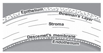
Anatomically the cornea has five layers
- Epithelium
- Bowman 's membrane
- Stroma (forms the bulk of corneal tissue)
- Descemet's membrane
- Endothelium
The cornea is avascular and has no lymphatics. Nerve supply to the cornea is from the branches of the trigeminal nerve.
The junction of the cornea and the sclera is called the limbus. Important angle structures lie here.
Uvea - The middle vascular layer
It is also called the uvea. This vascular layer supplies nourishment to the eyeball. It is composed of the iris, the ciliary body and the choroid. The uveal tract contains melanin- a pigment which determines the color of the eye.
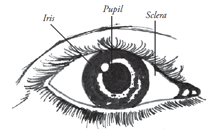
Iris
It is the most anterior portion of the uvea. It represents the pigmented moving diaphragm of he eye. It controls the amount of light entering the eye and is comparable to the aperture of the camera. The hole in the centre of the iris is the pupil. Pupil constricts in the light and dilates in the dark. Two muscles- the dilator and constrictor control this action. Constrictor muscles are innervated by parasympathetic fibers of the 3rd cranial nerve and the dilator muscles are innervated by sympathetic fibers from the superior cervical ganglion.
Ciliary body
It lies between the iris and choroid. It contains the ciliary muscle and ciliary processes. The ciliary muscle takes part in accommodation. With accommodation we change our focus at different distances. This is possible because of the simultaneous changes in the shape of the lens as the ciliary muscle contracts and relaxes.
The ciliary processes produce aqueous humor. Aqueous is a transparent fluid which nourishes ocular structures.
Aqueous humor
The aqueous humor is a clear, watery fluid, formed by the ultra filtration and secretion through the capillary process in the posterior chamber of the eye.
Circulation of aqueous
Pathway of aqueous humor drainage
Ciliary processes - posterior chamber - pupil - anterior chamber - trabecular meshwork - canal of Schlemm - collector channel - episcleral veins.
Note: Structures seen with a gonioscope while viewing angle structures - Schwab's line (end of Descemet's membrane), trabecular meshwork, scleral spur, ciliary body band, root of iris.
Choroid
It lies on the inner scleral surface. It nourishes the retina.
The receptor layer (retina)
This is the innermost layer of the eyeball. It is extremely thin (0.5mm) and transparent. It contains highly specialized visual receptors which help us see. It is comparable to the film of a camera. The retinal neurons transmit the picture through the optic nerve fibers to the brain for perception. The retina has ten layers:
- Retinal pigment epithelium
- Layer of rods and cones
- External limiting membrane
- Outer nuclear layer
- Outer plexiform layer
- Inner nuclear layer
- Inner plexiform layer
- Ganglion cell layer
- Nerve fiber layer
- Internal limiting membrane
The retinal receptors are divided into two main populations-the rods and the cones. The rods function best in dim light; the cones function best under daylight conditions. The cones are far fewer in number than the rods, numbering 6 million, whereas the rods number 125 million. Cones enable us to see small visual details with great acuity. Vision with rods is relatively poor. Color vision is totally dependent on the integrity of the cones. The cones form a concentrated area in the retina known as the fovea, which lies in the center of the Macula Lutea.
The junction of the periphery of the retina and the ciliary body is called the ora serrata.
Optic nerve
The optic nerve is the second cranial nerve situated in the posterior part of the globe. It transmits visual impulses from the retina to the brain. The head of the optic nerve is called the optic disc and is seen during ophthalmoscopic examination.
Visual pathway
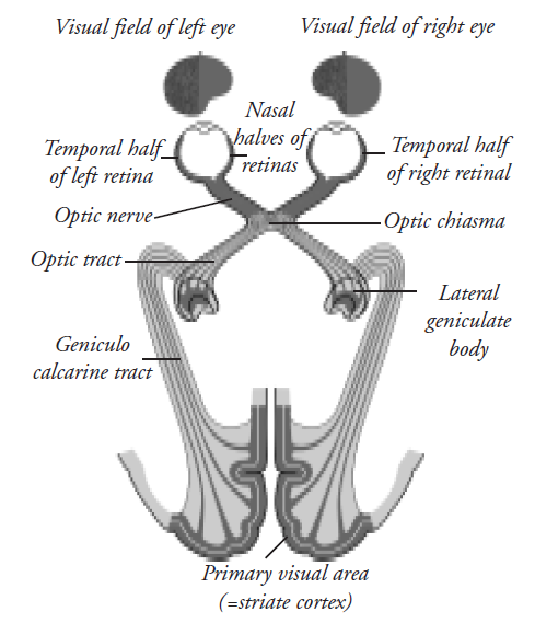
As the retinal fibers leave the optic nerves, half of them cross to the opposite side. This structure of mutual crossing is known as the optic chiasm. From the optic chiasm the crossed nasal fibers mix with the uncrossed temporal sector to form the optic tract. The optic tract continues towards a cell station in the brain called the lateral geniculate body. It is a relay station. From here fibers spread out in a fan shaped manner to reach their final destination the visual cortex (Fig. 4.5).
The lens
Lens which is proteinaceous and crystalline lens is a biconvex transparent structure. It lies behind the iris and in front of the vitreous. It is attached to ciliary muscles of the ciliary body by ligaments or fine threads, termed the zonules. Contraction of the ciliary muscle allows the zonules to relax. This in turn causes the lens to relax and become more convex. Thus it helps in accommodation.
The lens has an elastic envelope known as the capsule. The central portion of the lens is known as the nucleus. The nucleus is covered by cortex. The main function of the lens is to bend/refract light and to help in accommodation.
The Vitreous
The posterior segment of the eye. It prevents the eye from collapsing and provides support to the eye.
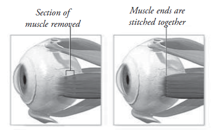
Extra ocular muscles
Each eyeball is controlled by six extra ocular muscles. There are four recti and two oblique muscles for each eye.
Note: The muscles are about 40mm long and about 10mm wide. They become tendinous 4-6mm from insertion.
Lacrimal system
Lacrimal system consists of
- Secretory portion
- Excretory portion
The main function of the lacrimal system is to produce tears for the lubrication and nourishment of the external eye. It also removes waste products (Fig. 4.6).
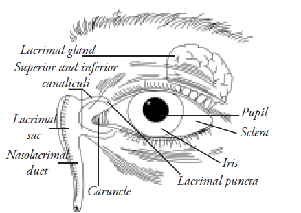
Secretory portion
The secretory portion consists of the main and accessory lacrimal glands. These glands produce the aqueous part of tears to keep the eye moist.
The main gland lies in the lacrimal fossa which is located at the anterio-lateral portion of the roof of the orbit. It opens by several ducts which pour tears into the superior fornix. This is responsible for reflex secretions.
The accessory glands of Krause and Wolf ring are located in the superior tarsal conjunctiva.
Excretory portion
This consists of
- Punctum
- Canaliculus
- Common canaliculus
- Lacrimal sac
- Naso lacrimal duct
Punctum
There are 2 puncta in each eye-the upper and the lower situated on the upper and lower eyelid margins about 5mm from medial canthus. Each punctum is situated on a lacrimal papilla.
Canaliculus
Each canaliculus has a 2mm vertical portion and an 8mm horizontal portion. The canaliculi are directed medially and meet to form a common canaliculus.
Lacrimal sac
The lacrimal sac is situated medially in the lacrimal fossa. The medial palpabral ligament lies anterior to it. It measures about 8-10mm.
Nasolacrimal duct
The nasolacrimal duct continues downward outward and laterally to end into the inferior meatus of the nose.
Tear film
The tear film resting on the corneal surface has three layers:
- Lipid or oil layer
- Lacrimal or aqueous layer, and
- Mucous or mucin layer
Functions of the tear film are to
- Carry bacteria-fighting compounds to the eye
- Carry nutrients to and waste products away from the eye
- Keep the eye moist
- Provide a smooth refracting surface
- Remove debris from the eye
Composition of tears
Tears contain almost 98% water. They are slightly alkaline due to the presence of chlorides and bicarbonates of sodium, potassium and calcium. They contain other constituents like urea, proteins, glucose, vitamin C and an antibacterial enzyme called lysozyme.
Functions of tears
The main functions of tears are:
- To keep the corneal and conjunctival surface moist and clean by mechanical flushing system.
- To provide nutrition to the cornea.
- To inhibit the growth of microorganisms
- To provide a regular corneal surface by filling interspaces between the epithelial cells.
Circulation of tears
Normally the amount of tear secretion is just sufficient to moisten the conjunctiva cul-de-sac. Almost 50% of the tear secretions are lost through evaporation. The rest are drained through the superior and inferior puncta medially by the blinking movements of the lids.
It is then sucked into the lacrimal sac and forced through the nasolacrimal duct into the nose during the act of blinking the eyes. During reflex irritation or emotional stimuli, excess of tears are secreted.
The amount of tear secretion can be measured by Schirmer's test. A strip of filter paper (5 by 35mm) is placed into the lower fornix for 5 minutes. At least 15mm of wetting indicates normal production.
Practical tips
Basal cell carcinoma, squamous cell carcinoma, melanoma and meibomian gland carcinoma may arise from the eyelids. These have to be recognized early and treated.
An extra row of eyelashes arising from the meibomian gland orifices is called dystichiasis. Misdirection of eyelashes which rub against the cornea is called trichiasis. In these conditions the eyelashes have to be removed (epilation).
Inward turning of the eyelid is called entropion.
- Outward turning of the eyelid is called ectropion.
- Loss of transparency of the lens or its capsule is called cataract and is seen commonly in middle - old age.
- Obstruction of the lacrimal drainage system causes epiphora while increased tear secretion is called lacrimation.
- Paralysis of the extra ocular muscle causes double vision (diplopia). This is commonly seen in 3rd, 4th, and 6th nerve palsies.
The visual system of the eye
The two eyes are the most important sensory organs of our body.
The human eye functions almost like a camera. It needs a transparent pathway of light from the outside world to be focused upon the retina (the innermost sensory layer of the eyeball - which acts like the film of a camera) to see images.
Parallel rays of light from a distant light source enter the eye through the clear pathway of cornea, aqueous humor, lens and the vitreous to form a sharp image upon the fovea in the retina. When the light falls upon the retinal receptors the rods and cones produce visual sensations due to photochemical changes in the pigment contents of the rods and cones, giving rise to electrical variations of charges, which are transmitted through the bipolar cells to the ganglion cells and ultimately via the visual pathway to the brain.
The visual cycle / Wald's cycle: When the retinal photoreceptors are exposed to light they undergo changes which can be transmitted to the brain. These chemical changes constitute the visual cycle.
Rods are made up of rhodopsin. (retinine + opsin = rhodopsin). This pigment, on exposure to light, gets bleached. This is the basic step of visual cycle.
The impulses from the retina travel along the visual pathway; that is: the optic nerve, optic chiasma, optic tract, lateral geniculate body, optic radiation and to the visual cortex of the brain. These impulses produce different sensations :
- Light sense: The rods are more sensitive to dim illumination, while the cones are sensitive to bright, day illumination
- Form sense: enables a person to perceive the shape of an object i.e., the visual acuity.
- Color sense: helps to distinguish different colors of different wavelengths of light.
In human beings the two eyes work together as if they were one and a single mental image is perceived. This is known as binocular singe vision.
Binocular vision is a unique feature of mammals and helps in specialized visual function.
It has three components
- Simultaneous perception
- Fusion
- Stereopsis
Simultaneous perception
Under normal conditions, the image of any object falls on the macula of each eye. There is simultaneous macular perception i.e. both the macula have the same image on them at one given time on looking in one direction.
Fusion
This is the ability to see two similar incomplete images simultaneously and interpret it as a single complete image
Stereopsis: depth perception
When binocular single vision is disturbed we see double. This is known as diplopia. By 6 months of age most of the binocular reflexes are well developed.
Pupillary reactions
When light is thrown on the eye, the pupils constrict. This is mediated by the afferent impulses traveling through the optic nerve and the efferent responses through the 3rd nerve.
These afferent responses bring about contraction of both the pupils due to the decussating of optic nerve fibres at the chiasma.
When the light stimulus constricts the pupil it is called a direct light response. The other pupil constricting is called consensual reflex.
Near response
When an object is brought close to the eyes, in order to focus it clearly on the retina, the eyes:
- Converge
- Accommodate
- The pupils constrict
This is called the near response
Summary
This unit has covered the development of the eye and different parts of the eye. The functions of all the parts of the eye are clearly described.
Key points to remember
- The eyeball is made up of three coats-the outer protective sclera, middle vascular choroids, inner neuronal retinal coat
- The transparent cornea is made up of 5 layers. Transparency of the cornea is maintained by its relative hydration. Cornea is the most important refracting media in the eye.
- The angle structures are important for drainage of aqueous humor and are important in the pathology of glaucoma
- The retina consists of 10 layers. The rods and cones are photoreceptors in the retina. Rods help in dim light and night vision and the cones for color vision and fine seeing.
- Lens is an important focusing mechanism of the eye. Ciliary muscle contraction relaxes the zonules resulting in increased refracting power of the lens. Thus the eye can see nearby objects.
Student exercise
A. Draw the diagram of eye and
- Describe the parts of the eye
- List the layers of the eyelid
B. True / False
- The cornea is the main refractive media of the eye. (True / False)
- The aqueous humour leaves the eye by filtering through the trabecular meshwork. (True / False)
- The rods are responsible for vision in dim light. (True / False)
- Loss of transparency of the lens is called cataract. (True / False)
- The accessory lacrimal glands are responsible for reflex tear secretion. (True / False)
- Relative afferent pupillary defect (RAPD) is tested by the swinging flashlight test. (True / False)
- Rods are responsible for colour vision (True / False)
C. Choose the correct answer
1. The orbicularis oculi is the muscle that
- Dilates the pupil
- Affects accommodation
- Closes the eyelids
- Constricts the pupil
2. The average size of the adult eyeball is
- 15mm
- 25mm
- 40mm
- 30mm
3. The meibomian gland is contained in
- Conjunctiva
- Muscle
- Tarsal plate
- Lacrimal system
4. Elevator muscle is supplied by (cranial nerve)
- 3
- 4
- 5
- 6
5. The aqueous is produced by
- Salivary gland
- Tarsal gland
- Lacrimal gland
- Ciliary body
All statements about tears are correct except
- Tears have a role in maintaining IOP
- Tears have bactericidal function
- Tears have cleansing function
- Tears have nutritional function
Answer the following
- Name the coats of the eyeball
- Which is the primary (most powerful) focusing structure of the eye?
- Name the parts of the vascular coat of the eye and the source of its dark color
- How does the pupil control the amount of light entering the eye?
- Trace the flow of aqueous in the eye
- Name the structures seen in the angle of the eye
- Name the receptors in the retina and their function
- List the extra ocular muscles, their origin, insertion, action and nerve supply
- What is accommodation? Name the structures that are needed for the eye to see nearby things clearly
- What is aqueous humor? Describe its functions.
- Name the layers of the tear film and the functions of tears
- Name the pupillary reactions. Describe the method to test direct and consensual pupillary reflex
Practical skill student's exercise / activity
- Label the various parts of the eye in the charts provided.
- Collect information on the mechanism of aqueous humor production and drainage in detail and prepare a chart for the same.
- Enumerate various refractive media of the eye. Name the most important one and its refractive index.
- Describe the pupillary pathway with well illustrated diagrams.